Reading residual blood flow in a blocked brain artery from a routine CT scan
Published in Stroke, 2025 · Summary posted 5 July 2026
In plain language
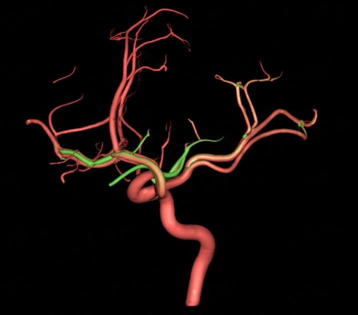
During a stroke, one of the brain's arteries can become blocked by a clot. Whether a small amount of blood still manages to flow forward past that blockage (called antegrade flow) can influence how well a person recovers.
This study tested whether an automated software tool called vascular territory mapping, which works out which artery is supplying each part of the brain using a standard CT perfusion scan, can help identify that residual forward flow. Using detailed 3D CT angiography as the benchmark and patients drawn from an international stroke imaging registry, we found the software was a useful indicator of antegrade flow in people with a proximal (M1) blockage.
Why it matters
Spotting preserved forward flow quickly, from imaging that is already part of routine stroke assessment, can add to a treatment team's picture of each patient. It also supports the broader goal I work on: building automated tools that read stroke scans faster and more consistently, so the right patients are identified sooner.
The paper
This is a summary of published research written for general readers, not medical advice. If you or someone you care for has symptoms of stroke, call 000 (in Australia) immediately. For questions about your own health, please speak with your doctor.
← Back to all research summaries